
A Clinician's Guide to the Grading Muscle Strength Scale
Team Meloq
Author
At the heart of any physical assessment lies the need to measure a person's functional power. For this, clinicians rely on the grading muscle strength scale, a simple yet effective tool for quantifying muscular output. The most common system, the Medical Research Council (MRC) scale, uses a straightforward 0-to-5 rating to create a universal language for healthcare professionals tracking patient progress and documenting changes in strength.
The Clinical Standard for Muscle Testing
When a physiotherapist, physician, or athletic trainer needs to assess muscle function, they turn to a time-tested method called Manual Muscle Testing (MMT). The MRC scale, also known as the Oxford Scale, is the backbone of this process. It provides a quick, hands-on way to grade a muscle's ability to contract and generate force against resistance.
This scale can be viewed as a standardized ruler for muscle power. Every number on the 0-to-5 scale corresponds to a specific level of function, starting at Grade 0 (a complete lack of visible or palpable muscle contraction) and progressing to Grade 5 (normal strength against maximal resistance).
This systematic approach is critical for several clinical purposes:
- Monitoring Recovery: It is used extensively to track progress following surgery, injury, or a neurological event like a stroke.
- Diagnosing Conditions: The scale helps identify patterns of weakness that can point toward specific nerve injuries or muscle diseases.
- Guiding Rehabilitation: The grades allow clinicians to design effective, targeted exercise programs based on a patient’s current strength levels.
This visual guide breaks down the basic hierarchy of muscle movement that the scale assesses.
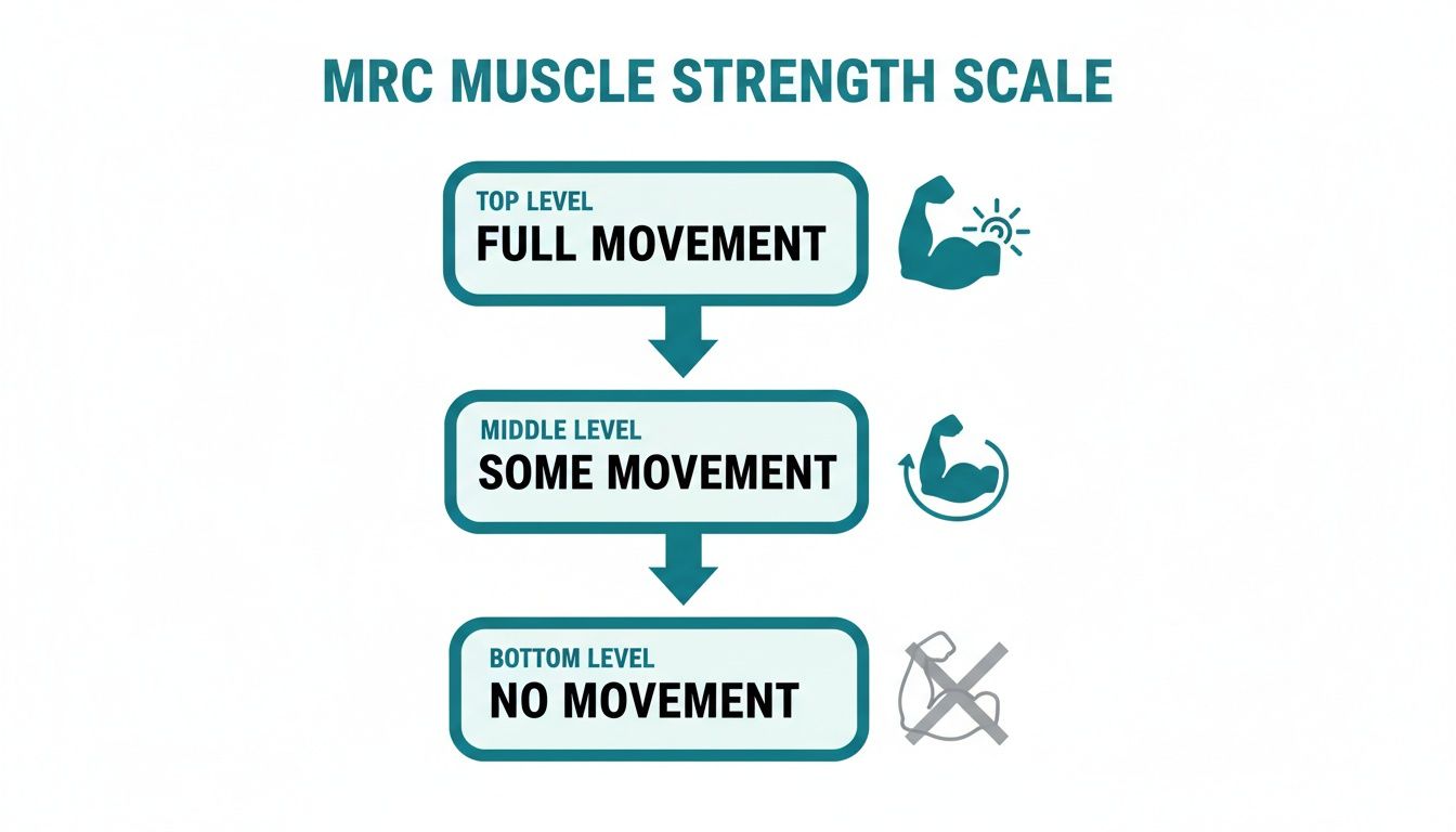
As shown, the core concept is a logical progression from no discernible muscle activity to full, functional movement.
Why It's So Widely Accepted
The MRC scale's prominence in clinical settings is well-established. It is recognized as a widely accepted method for grading muscle strength in both the upper and lower extremities.
The scale's integration into routine neurological exams is extensive across the US, Europe, and other regions, making it a key component in diagnosing conditions ranging from stroke to various myopathies. You can learn more about the clinical application of muscle testing and its foundational role in physical assessment.
This broad acceptance means that a "Grade 4 quadriceps" has a consistent meaning for a physiotherapist in London and a sports physician in Los Angeles. Such consistency is vital for clear communication across different healthcare disciplines, ensuring patients receive seamless care as they move between specialists.
Decoding the MRC and Oxford Muscle Strength Scale
To understand how the scale works in practice, it is helpful to see each grade broken down with a clear example. This table clarifies the clinical definitions.
| Grade | Official Description | Simple Explanation | Example |
|---|---|---|---|
| 0 | No palpable muscle contraction | The muscle shows no sign of activity. Nothing happens when the patient tries to move. | A patient after a severe stroke cannot produce any flicker of movement in their biceps. |
| 1 | Flicker or trace of contraction | You can see or feel a tiny twitch in the muscle, but it is not enough to move the limb. | The same patient can now create a tiny, visible twitch in their biceps, but their arm does not bend. |
| 2 | Active movement, with gravity eliminated | The person can move the limb through its full range of motion, but only if gravity is not a factor (e.g., lying on their side). | Lying on their side, the patient can now slide their arm across a table to bend their elbow. |
| 3 | Active movement against gravity | They can move the limb through its full range of motion against the force of gravity, but cannot overcome any additional resistance. | The patient can bend their elbow fully while sitting up, but their arm gives way if any external pressure is applied. |
| 4 | Active movement against gravity and some resistance | The muscle can move against gravity and resist some pushback from the examiner, but it is not at full strength. | The patient can bend their elbow against gravity and hold it against moderate pressure, but "breaks" with more forceful resistance. |
| 5 | Normal power. Active movement against full resistance | The muscle has normal strength and can hold its position against the examiner’s maximum resistance. | The patient can hold their biceps contraction strong and steady against a clinician's best effort to straighten their arm. |
Ultimately, the scale's simplicity is one of its greatest assets. It serves as a foundational skill for students, a quick reference for seasoned clinicians, and a helpful guide for patients seeking to understand their own recovery journey.
Born From the Battlefield: The Wartime Origins of a Clinical Standard
To fully appreciate why the muscle strength grading scale is so fundamental, one must look back at its origins. This tool was not developed in a quiet laboratory but was forged out of the urgent needs of World War II.
At that time, clinicians faced a significant challenge: how to consistently evaluate soldiers with peripheral nerve injuries. Without a shared system, one physician’s "weak" was another’s "recovering," leading to inconsistent care. A simple, repeatable method was needed to track recovery and make critical decisions on the battlefield and in military hospitals.
A Tool Forged in Conflict
The breakthrough came from the UK's Medical Research Council (MRC). They were tasked with creating a universal framework for manual muscle testing that could be learned and applied consistently.
The result was the Oxford Muscle Strength Scale, now known universally as the MRC scale. It was first published in a 1943 pamphlet titled 'Aids to the Investigation of Peripheral Nerve Injuries,' created specifically to help medics assess nerve damage in wounded soldiers. You can trace its journey from a wartime tool to a modern standard at Physiopedia.
This wartime innovation gave clinicians a clear, 0-to-5 grading system. It was straightforward enough for any trained professional to use, ensuring that an assessment in a field hospital in France would mean the same thing as one in a recovery ward in England. This consistency was a game-changer for tracking nerve regeneration and guiding rehabilitation for service members.
The scale’s brilliant simplicity is why it outlasted the war and transitioned so smoothly into civilian medicine. It quickly became a cornerstone of physiotherapy, neurology, and rehabilitation globally. Decades later, its principles were expanded to create tools like the MRC Sum-Score (MRC-SS), which is now indispensable for diagnosing complex conditions like ICU-acquired weakness.
This journey—from a tool of military necessity to a benchmark of modern clinical practice—demonstrates the lasting power of an elegant, practical solution to a complex problem.
Performing Manual Muscle Testing with Precision
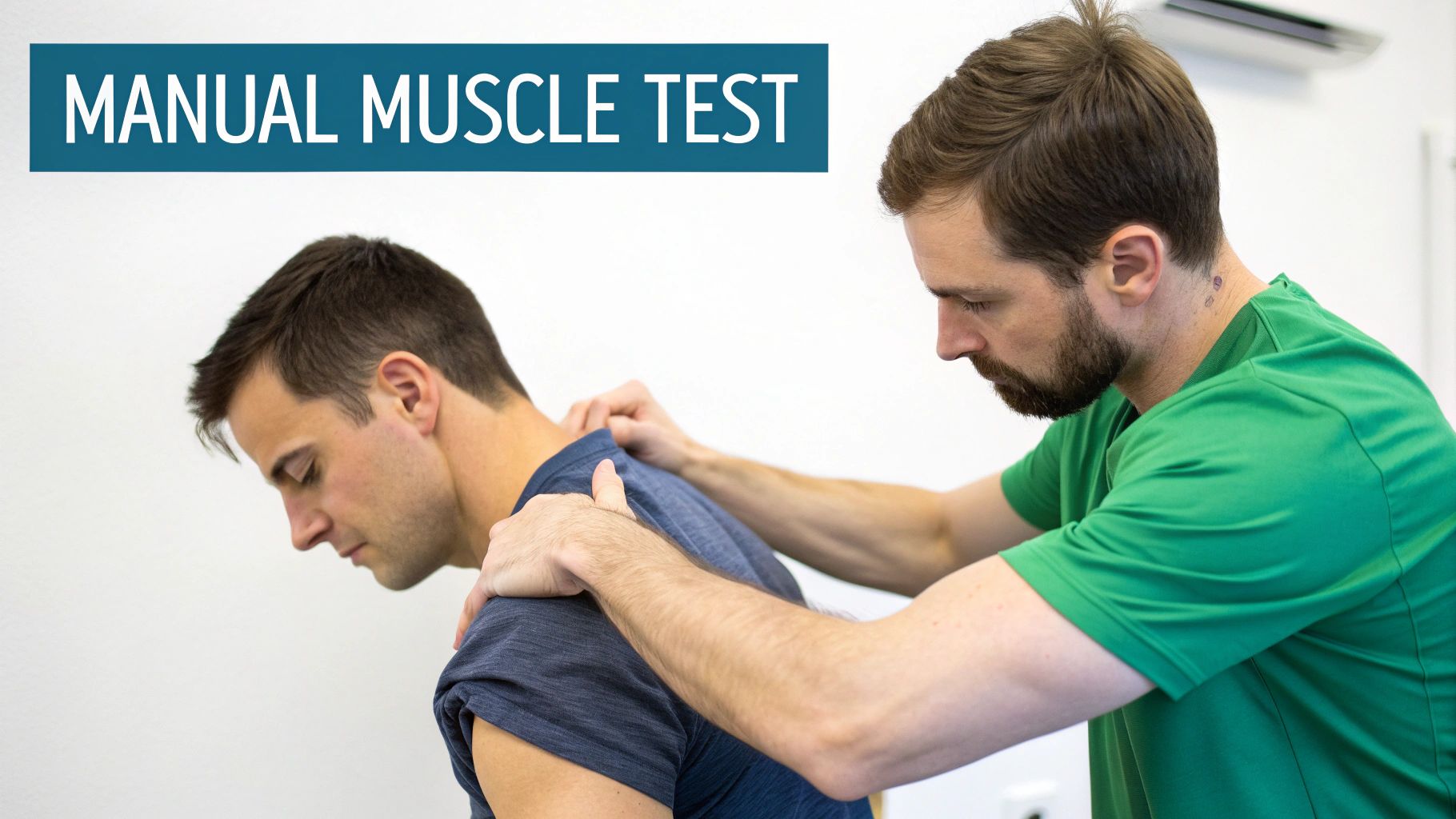
Knowing the muscle strength grades is one thing; applying them with skill is another. Mastering Manual Muscle Testing (MMT) is a hands-on skill that requires precision. Proper technique ensures the grades recorded are reliable and repeatable, providing a true snapshot of a patient's functional strength.
An experienced clinician develops a feel for subtle differences in muscle activation and resistance, but that intuition is built on a solid foundation of standardized procedure. Consistent technique is what separates a guess from an assessment. It is how one reliably differentiates between the flicker of a Grade 1 contraction and the observable movement of a Grade 2, or a subtle Grade 4 weakness from solid Grade 5 strength.
The Foundation of a Reliable Test
To achieve consistent results, every MMT session should follow a structured approach. This involves more than just pushing on a limb; it is about systematically isolating the target muscle to remove as many variables as possible. The goal is to ensure the test reflects that muscle's true capacity.
A few core principles are non-negotiable for a valid test:
- Optimal Positioning: Always place the patient in a position that isolates the target muscle. This often means using specific postures to prevent other muscles from compensating for weakness.
- Clear Instructions: Use simple, direct language. Tell the patient exactly what to do, and coach them to "hold" their position as you apply resistance.
- Proper Stabilization: You must stabilize the body part just proximal to the joint being tested. For example, when testing shoulder abduction, stabilizing the scapula is essential. If not, the entire shoulder girdle can elevate, giving a false impression of deltoid strength.
Against Gravity vs. Gravity-Eliminated Positions
One of the most critical decisions during testing is choosing the right position, which is guided by the patient's initial ability.
As a general rule, always start with an against-gravity test. If a patient can move through their full range of motion against gravity, their strength is at least a Grade 3. Only if they are unable to do so should you switch to a gravity-eliminated position to differentiate between a Grade 1 and Grade 2.
Let's use the shoulder abductors (deltoid muscle) as an example:
- Against-Gravity (Grade 3+): The patient sits upright and attempts to lift their arm straight out to the side. If they complete the full motion, you then apply resistance to determine if they are a Grade 4 or Grade 5.
- Gravity-Eliminated (Grade 2): If they could not lift their arm, have them lie on their back. Now, ask them to slide their arm out to the side along the table. If they can do this, you are looking at Grade 2 strength.
This methodical approach is essential for accurate grading. This systematic process transforms a subjective "feel" into a dependable clinical measurement, allowing clinicians to apply the grading scale with confidence.
Navigating the Limitations and Common Pitfalls
While the muscle strength grading scale is a workhorse in clinical practice, it is important to acknowledge its limitations. Its greatest strength—simplicity—is also the source of its biggest weakness: subjectivity.
What one clinician considers "maximal resistance" for a Grade 5 test might differ from a colleague's perception. This inter-rater variability can complicate the process of tracking a patient's progress over time.
This subjectivity is particularly pronounced in the higher grades. The functional gap within Grade 4, defined as movement against "some" resistance, is notoriously wide. A patient might progress from barely resisting a push to demonstrating near-full strength, yet their official score could remain at Grade 4. This can mask subtle but clinically meaningful improvements.
Understanding these constraints helps clinicians use the scale more effectively and recognize when to incorporate objective tools for a clearer picture.
The Challenge of Differentiating Higher Grades
Differentiating the upper tiers of strength is arguably the most difficult aspect of using the MRC scale. Distinguishing between a strong Grade 4, a 4+, or a true Grade 5 often depends as much on the examiner's strength and experience as on the patient's actual power output.
This is a well-documented psychometric issue. One key study found disordered thresholds in a staggering 74-79% of muscles tested, meaning clinicians consistently struggled to differentiate between adjacent grades (1). The research highlighted that distinguishing Grade 4 from 4+ or 5 was particularly unreliable. The authors even proposed a modified system to improve precision. For those interested, you can explore the full research findings on the MRC scale's psychometric properties.
Common Pitfalls and Influencing Factors
Beyond subjectivity, several other factors can skew the results of a manual muscle test, leading to an inaccurate grade. Awareness of these common errors is the first step toward more reliable assessments.
- Patient-Related Factors: Pain is a significant confounder. A patient may possess the underlying strength for a Grade 5, but if the test is painful, they will instinctively inhibit their effort. This results in a lower score that does not reflect their true capacity. Fatigue, motivation, and comprehension of instructions can also influence the results.
- Tester-Related Factors: The primary issue here is inconsistent force application. Pushing too quickly, too slowly, or at an improper angle can change the outcome. Another classic mistake is poor stabilization, which allows other muscles to compensate and create a false impression of strength in the target muscle.
To help achieve more reliable results, the following table outlines common pitfalls and how to avoid them.
Common Pitfalls in Manual Muscle Testing and How to Avoid Them
| Common Pitfall | Impact on Assessment | Practical Solution |
|---|---|---|
| Inconsistent Resistance | Applying force too quickly, slowly, or unevenly leads to unreliable and non-repeatable results. | Apply resistance smoothly and gradually over 3-5 seconds. Always aim to test at the same point in the range of motion. |
| Poor Stabilization | Allows for compensation from other muscle groups, giving a false high score for the muscle being tested. | Use your free hand (or body) to firmly stabilize the joint proximal to the one being tested. Ensure the patient is in a stable, supported position. |
| Testing Through Pain | Pain can cause reflex inhibition, leading to a score that reflects pain tolerance, not true muscle strength. | Modify the test to be pain-free if possible. If not, document that the test was limited by pain (LBP) and consider other assessment methods. |
| Vague Verbal Cues | If the patient doesn't understand "hold, don't let me move you," their effort will be suboptimal and inconsistent. | Use simple, direct, and consistent commands. Demonstrate the movement first. Encourage maximal effort throughout the test. |
| Ignoring Fatigue | Testing a long series of muscles without breaks can lead to lower scores on later tests due to general fatigue. | Plan your testing sequence logically. Allow for brief rest periods, especially when testing multiple large muscle groups or a weak patient. |
| Grade 4 "Fuzziness" | The wide range of Grade 4 makes it difficult to track subtle but meaningful progress within this grade. | Use "+" and "-" modifiers consistently and document your specific rationale. Supplement with objective tools like dynamometry for patients in this range. |
By keeping these points in mind, clinicians can improve their assessment quality. The goal is to build consistency by using established protocols, comparing findings to normative data where available, and maintaining meticulous records.
Streamlining the documentation of these detailed muscle assessments can be aided by tools like medical voice charting. By sidestepping these common pitfalls, you can navigate the scale's limitations and deliver a more accurate and defensible assessment of a patient's true functional capacity.
Moving Beyond Manual Grades With Objective Data
Manual muscle testing is a foundational clinical tool, but its reliance on subjective "feel" can mask subtle yet critical changes in strength, especially in the higher grades. To track progress with greater precision, clinicians are increasingly turning to technology that provides objective, quantifiable data.
This is not about replacing the trusted grading muscle strength scale, but rather enhancing it. By layering hard numbers onto clinical assessments, we can build a more complete, defensible, and insightful strength profile for every patient.
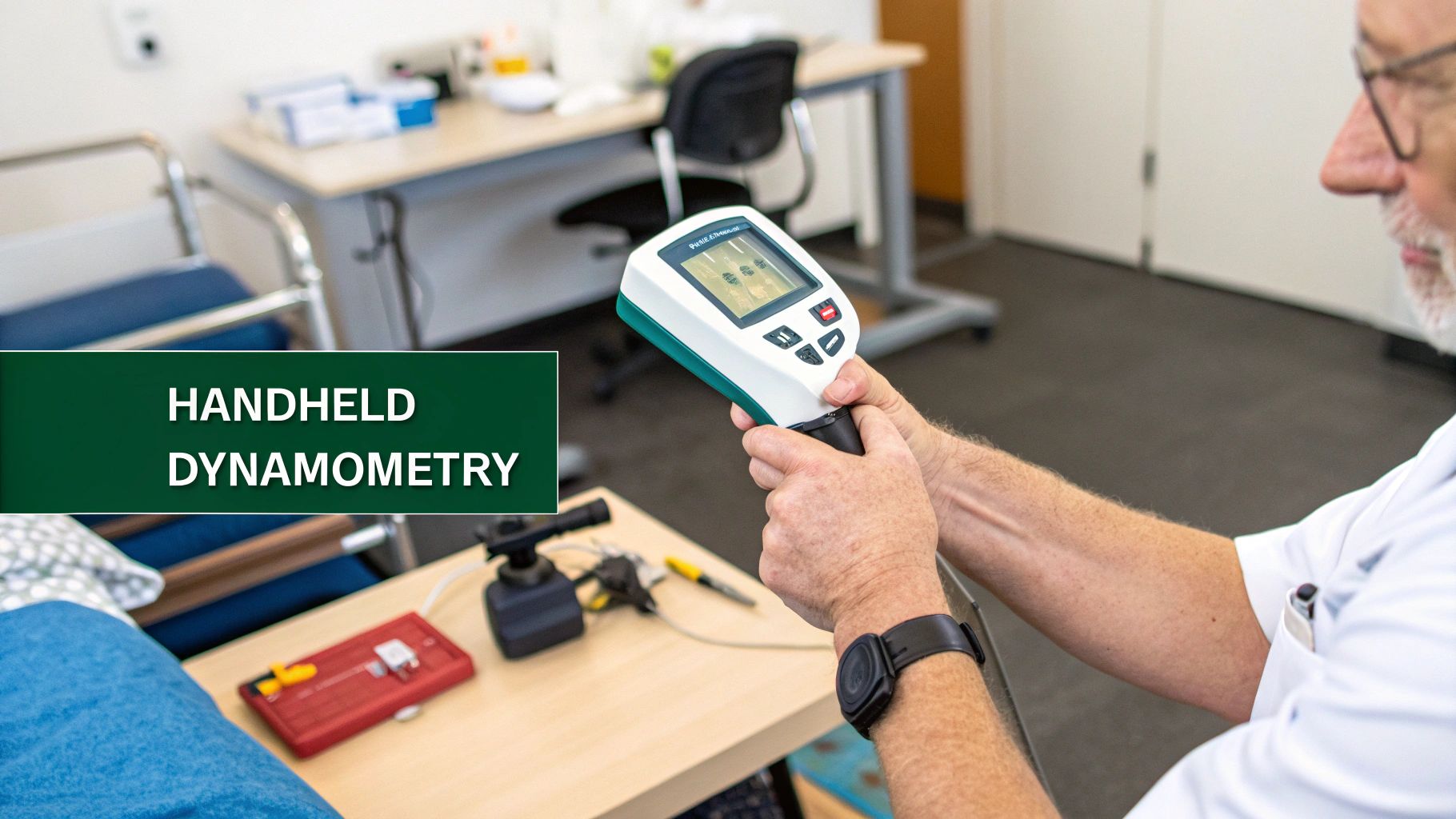
Capturing Force With Handheld Dynamometry
The most direct way to address the guesswork in Grade 4 and Grade 5 assessments is with handheld dynamometry. These portable devices measure the actual force a muscle can generate, providing a clear number in kilograms, pounds, or Newtons. This removes the ambiguity of deciding if a patient is providing "some" or "full" resistance.
Instead of just recording "Grade 4," you can document that a patient’s quadriceps produced 25 kg of force. A month later, a reading of 30 kg represents undeniable, measurable progress that MMT alone might have missed entirely.
These concrete numbers are invaluable for tracking small but significant improvements over time. The use of such objective tools is particularly important when assessing diverse populations, including those who may receive support through grants for people with disabilities, where precise tracking is key. To better understand how these devices function, one can explore the different types of dynamometers used for muscle testing.
Complementing, Not Replacing, the MRC Scale
It is important to understand that there is no direct formula to convert a dynamometer reading into an MRC grade. That is not the purpose. Instead, these tools work in tandem to create a richer clinical picture.
- MRC Grades 0-3: For identifying a flicker of contraction or the ability to move against gravity, standard MMT is highly effective. It is fast, efficient, and provides the necessary information at this stage.
- MRC Grades 4-5: This is where dynamometry excels. It provides the objective data needed to differentiate between "strong" and "truly strong," and to track changes within this broad range.
The Gold Standard: Isokinetic Dynamometry
For the highest level of precision, particularly in high-level sports performance and research settings, isokinetic dynamometry is considered the gold standard. These are large, computer-controlled machines found in laboratories that measure muscle force at a constant speed through an entire range of motion.
While not practical for most clinics, they represent the upper end of the strength assessment spectrum. This illustrates the progression in the field—from a quick bedside MMT to highly detailed, data-driven analysis, reflecting the push toward more objective, evidence-based practices in rehabilitation and performance.
A Checklist for Reliable Strength Assessments
Understanding the theory behind muscle testing is one thing; applying it consistently is another. To get the most out of the grading muscle strength scale, a systematic approach is essential.
The following points can serve as a pre-test checklist. This is about building a solid, repeatable process to turn subjective feelings into meaningful, defensible data.
This checklist distills the core lessons of this guide into simple, actionable steps for any clinical or field setting, aiming to reduce variability and guesswork for more trustworthy results.
Standardize Your Testing Protocol
First, create a consistent routine for every muscle group you test and adhere to it strictly. This means using the exact same patient positioning, stabilization techniques, and point of resistance application every time.
Documenting this protocol is crucial. It ensures that you, and anyone else in your practice, are measuring the same parameter in the same way from one visit to the next. Without this level of standardization, it is difficult to know if a change in grade is due to actual patient progress or a variation in testing methods.
Use Consistent Verbal Cues and Instructions
How you instruct a patient can dramatically alter their effort and the result. Vague or changing commands introduce a significant variable.
Adopt a script and stick to it. Simple, direct phrases like, "Hold this position. Don't let me move you," are clear and effective. Coaching the patient to ramp up to their maximum effort is also key to assessing their true strength potential.
Ensure Meticulous Stabilization
A common error in manual muscle testing is allowing patient compensation. If you are testing shoulder abduction but fail to stabilize the scapula, the patient may hike their entire shoulder girdle. This gives a false impression of deltoid strength when it is actually a compensatory movement.
Always secure the body part just proximal to the joint being tested to isolate the target muscle and ensure it is the primary muscle working.
Test Bilaterally for Comparison
Make it a habit to test the unaffected side first. This is important for two reasons. First, it provides an immediate baseline of that patient’s "normal" strength. Second, it serves as a rehearsal, allowing the patient to understand the task before you test the affected side.
This context is invaluable for interpreting your findings. To take this a step further with objective numbers, it helps to understand what force measurement is and how tools like dynamometers can complement these hands-on skills.
Document All Influencing Factors
Your clinical notes should tell the full story, not just the final grade. If a patient's effort was clearly limited by pain, or if they were fatigued or unmotivated, this should be documented.
This context is critical for anyone else reading your notes and for tracking progress over time. It makes your clinical picture more complete, defensible, and truly justifies the grade assigned on the muscle strength scale.
Answering Your Questions About Muscle Strength Grading
As you use muscle grading scales, a few common questions often arise. Addressing some of the most frequent inquiries can help navigate real-world scenarios with more confidence.
What Is the Biggest Limitation of the MRC Scale?
The primary limitation is subjectivity, particularly at the higher end of the scale. The distinction between a Grade 4 ("movement against some resistance") and a Grade 5 ("normal power") can be ambiguous and may vary between clinicians—or even for the same clinician on different days.
Furthermore, Grade 4 covers a wide functional range. A patient who can barely resist a light touch and an athlete who is only slightly weaker than their uninjured side could both fall into this category. This makes it difficult to track small yet clinically significant improvements in strength, which is a key reason why objective tools like handheld dynamometry are valuable complements to the MRC scale (1).
Can This Scale Be Used on Unconscious or Sedated Patients?
Not in its traditional form. The standard MRC scale requires active patient participation and the ability to follow commands, so it is not suitable for individuals who are unconscious or heavily sedated.
However, in critical care settings, clinicians use adapted methods to assess muscle function. This may involve observing reflexive movements or using a peripheral nerve stimulator to elicit a muscle contraction, which can then be graded on a simpler scale. The MRC Sum-Score is a critical tool for diagnosing ICU-acquired weakness once a patient is sufficiently awake to participate in testing (2).
How Do I Choose Between Testing Positions?
Let the patient's ability guide your decision. As a rule of thumb, always begin by asking the patient to perform the movement against gravity.
If they can move through the full range of motion, their strength is at least a Grade 3. From there, you can apply resistance to determine if they are a Grade 4 or 5. If they cannot complete the movement against gravity, the next step is to reposition them into a gravity-eliminated plane. If they can complete the full range of motion in this new position, their strength is graded as a 2.
For clinicians ready to move beyond subjective assessments and capture objective, repeatable data, Meloq offers a suite of digital measurement tools. Our EasyForce dynamometer provides precise force readings to complement your manual muscle testing, helping you track patient progress with undeniable accuracy. Discover how to enhance your clinical practice at https://www.meloqdevices.com.
References
- Bohannon RW. Manual muscle testing: does it meet the standards of an adequate screening test? Clin Rehabil. 2005;19(6):662-7.
- Vanpee G, Segers J, Van Mechelen H, Wouters P, Van den Berghe G, Hermans G. The MRC sum score: a simple method to quantify muscle weakness in the intensive care unit. J Thorac Dis. 2011;3(1):31-6.

Featured Product
EasyForce Digital Dynamometer
Handheld muscle strength testing with 99% accuracy. Used in 40+ peer-reviewed studies.
Learn More